Palmar angle describes the orientation of the solar margin of the distal phalanx relative to the ground in the standing horse. Although palmar angle is frequently discussed in the context of loading and locomotion, it is fundamentally a physiological relationship that emerges from the spatial organisation, suspension, and connective tissue integration of the equine digit. Understanding the contribution of the digital cushion to palmar angle therefore requires precise clarification of its three dimensional position, its biological functions, and its mechanical integration with the distal limb. Many contemporary debates surrounding the digital cushion arise not from disagreement about its importance, but from oversimplified anatomical models that attribute to it roles that are not supported by histology or gross anatomy.

Spatial anatomy of the digital cushion
Gross anatomical and histological studies consistently describe the digital cushion as a specialised caudal structure occupying the space between the medial and lateral ungual cartilages at the back of the foot. It is composed of fibro elastic connective tissue with variable adipose content and regionally fibrocartilaginous characteristics, and it blends dorsoproximally with dense collagenous tissue that is continuous with the deep digital flexor tendon and the podotrochlear apparatus (Bowker et al., 1998; Bowker, 2003). Pollitt similarly describes the ungual cartilages as attaching along the semilunar line of the distal phalanx and projecting axially into the digital cushion over the bars, framing the cushion as a caudal mass rather than a broad plantar structure beneath the body of the distal phalanx (Pollitt, 1996; Pollitt, 2010).
This three dimensional arrangement is frequently misinterpreted when viewed through isolated mid sagittal sections of the foot. Such sections can create the visual impression that the digital cushion lies extensively beneath the distal phalanx. However, transverse and parasagittal histological sections demonstrate that the greatest volume of the digital cushion lies caudal to the palmar processes of the distal phalanx, filling the heel region between the ungual cartilages and underlying the frog and heel bulbs. Beneath the dorsal and central portions of the distal phalanx, the tissues interposed between bone and ground are primarily solar corium and sole producing epidermis, not digital cushion tissue. The solar corium is tightly adherent to the solar surface of the distal phalanx and constitutes a distinct anatomical compartment from the digital cushion, which represents a modification of the caudal subcutis (Pollitt, 1996).
The digital cushion is not, however, confined exclusively to a position behind the distal phalanx. Anatomical descriptions of the frog digital cushion complex demonstrate a thinner cranial extension that runs forward beneath the palmar portion of the distal phalanx, ventral to the deep digital flexor tendon and proximal to the frog apex. O’Grady and Poupard describe this extension as mirroring the frog and lying along the palmar solar surface of the distal phalanx in the region of the flexor surface and palmar processes (O’Grady and Poupard, 2003). This cranial extension establishes anatomical proximity between the digital cushion and the palmar aspect of the distal phalanx, but it represents a transitional zone rather than the primary mass of the structure.
Importantly, equine anatomical and histological studies do not quantify the proportion of the digital cushion lying beneath the distal phalanx in numerical terms. Instead, they describe spatial relationships, attachments, and regional transitions. What emerges clearly is a qualitative partition. The bulk of the digital cushion volume is caudal, situated between the ungual cartilages and above the frog and heel bulbs, while a thinner extension lies beneath the palmar solar region of the distal phalanx. The digital cushion is therefore not a uniform pad beneath the body of the distal phalanx, but a caudal wedge shaped mass with a forward extension under the palmar aspect.
Biological function and tissue specialisation
The geometry and composition of the digital cushion have direct implications for its function. Histological composition and vascular architecture indicate that its primary biological roles are haemodynamic regulation and shock dispersion rather than static skeletal support. Bowker proposed that the digital cushion, together with the ungual cartilages and frog, forms a central component of the venous pump of the equine foot, assisting venous return through deformation driven by both internal skeletal motion and external ground interaction (Bowker et al., 1998; Bowker, 2003).
This haemodynamic role is supported by histological evidence demonstrating that the digital cushion is well vascularised. Detailed mapping studies describe a dense network of veins, venous plexuses, and arteriovenous anastomoses within the digital cushion, with regional variation in vascular density and close integration with the vasculature of the ungual cartilages and frog corium. Such a vascular arrangement is incompatible with the notion of a poorly perfused tissue and supports the interpretation of the digital cushion as an active participant in distal limb circulation.
A common misconception arises from conflating vascularity with regenerative capacity. The digital cushion is mesenchymal connective tissue composed of fibro elastic tissue, adipose tissue, and regionally fibrocartilage. It does not possess a germinative epidermal layer. Unlike the hoof wall, sole, or frog horn, which are produced by specialised germinative epithelia, the digital cushion does not grow horn and does not regenerate through epidermal proliferation. While it is well perfused, its capacity for structural regeneration is limited once its internal architecture is disrupted or replaced by fibrosis. Changes in digital cushion quality over time therefore reflect adaptive remodelling rather than true regeneration. This distinction explains why the digital cushion can be healthy, robust, and well perfused, yet slow or unable to recover once significantly compromised.
Mechanical behaviour and deformation
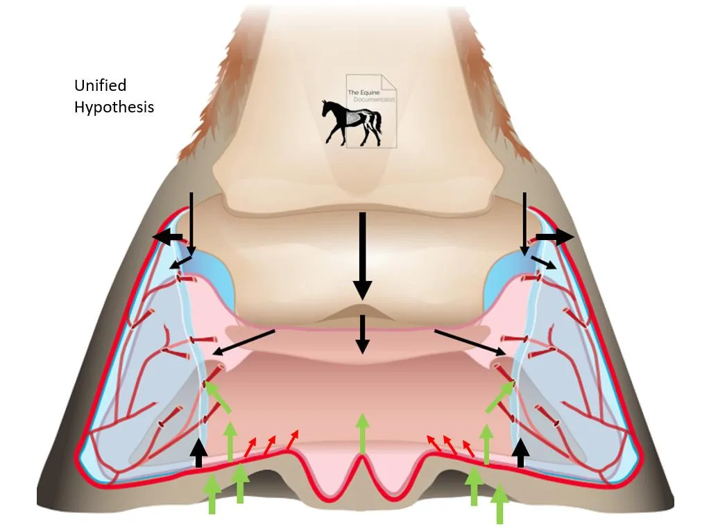
Mechanically, the digital cushion is designed to be deformed rather than to resist deformation. It accommodates descent of the middle phalanx and changes in distal interphalangeal joint posture, acting as a compliant interface within the caudal foot. It is also displaced by ground reaction forces transmitted through the frog and frog stay apparatus. These deformation patterns reflect its integration within a tightly bound fascial and connective tissue network that encompasses the entire caudal hoof, including the digital cushion, frog, ungual cartilages, podotrochlear apparatus, and associated ligaments and tendons. The dense connective tissue composition of the digit means that all components are mechanically coupled, and changes in one element propagate through the system rather than remaining isolated.
In this context, the digital cushion can reasonably be described as part of a non linear mechanical system. As deformation increases, progressively stiffer connective tissue elements and geometric constraints may be engaged, giving rise to a rising resistance to compression. This behaviour is consistent with viscoelastic damping and progressive stiffness, but it should not be simplified into a single stacked model in which the navicular bone directly compresses the digital cushion from above while ground reaction forces compress it from below. The navicular bone primarily interacts with the deep digital flexor tendon and its bursa, and its motion is constrained by joint geometry and tendon mechanics. Compression of the caudal hoof arises from the combined effects of phalangeal posture, tendon forces, frog ground interaction, and confinement by the ungual cartilages and hoof capsule.
Palmar angle as a system outcome
Within this integrated anatomical and mechanical framework, palmar angle emerges as a product of biotensegrity and morphology rather than the influence of any single structure. The distal phalanx is suspended within the hoof capsule by the laminar attachment, and its orientation is constrained proximally by the distal interphalangeal joint and the posture of the middle phalanx. External hoof capsule geometry, particularly heel to toe height ratios, directly influences palmar angle because altering capsule proportions changes the spatial relationship between the distal phalanx and the ground. Trimming or shoeing interventions that modify heel height reliably alter palmar angle, even when such changes increase tension within the deep digital flexor tendon.
The role of the deep digital flexor tendon in influencing palmar angle is well established, particularly in flexural deformities such as club foot, where increased resting tension within the tendon is associated with increased palmar angle (Redden, 2003; Dyson, 2011). However, the observation that palmar angle can be modified through hoof capsule geometry even when deep digital flexor tendon tension increases demonstrates that no single tissue dictates the outcome. Instead, palmar angle reflects the balance of constraints imposed by laminar suspension, joint posture, tendon tension, capsule geometry, and the connective tissue continuum of the digit.
The digital cushion participates in this balance not as a rigid support beneath the distal phalanx, but as an adaptive, deformable component within the caudal hoof complex. Its continuity with the podotrochlear apparatus and deep digital flexor tendon provides an anatomical basis for the strong correlation observed between phalangeal alignment, tendon tension, and digital cushion morphology. In this relationship, the digital cushion largely adapts to the posture and constraints of the digit rather than dictating them. A healthy, robust, and well perfused digital cushion strengthens the biotensegrity of the caudal hoof complex by improving deformation control, haemodynamic efficiency, and force distribution, thereby increasing the tolerance of the system to imposed geometry and posture.
Reconciling Mechanical and Haemodynamic Interpretations of the Digital Cushion
If personal positions are set aside and the question is reduced to what published anatomy and histology actually support, the evidence points to a clear both-and conclusion rather than an either-or dispute.
First, the digital cushion functions as a viscoelastic, non linear damping structure. It sits within the force path between the middle phalanx, the podotrochlear apparatus, the frog, and the ground, and it deforms in response to changes in phalangeal posture and external forces. Its fibro elastic and regionally fibrocartilaginous composition is consistent with a structure designed to absorb energy, dissipate forces, and exhibit progressive stiffness with increasing deformation.
Second, there is credible anatomical and histological evidence that the caudal hoof contains a substantial microvascular network, and that deformation of the caudal tissues can plausibly contribute to venous return and flow regulation. This remains true even if the digital cushion itself is not a large blood reservoir.
Vascularity of the digital cushion
Histology does not support the claim that the digital cushion has a poor blood supply as a general rule. A detailed histologic mapping study of the digital cushion in sound Quarter horses quantified vessel number, lumen diameter, and wall thickness, and reported a large number of vessels, particularly in the proximal regions of the cushion. That same study demonstrated clear regional variation in vascular density, with greater vascularity proximally than distally.
Importantly, the authors explicitly framed the digital cushion as having both mechanical and haemodynamic relevance, stating that the extensive vasculature observed provides evidence for a role in blood flow regulation alongside absorption and dissipation of applied forces. This directly contradicts the notion that the digital cushion is functionally avascular.
The correct anatomical correction, therefore, is straightforward. The digital cushion is not avascular. It contains measurable vascular structures. Apparent vascularity varies by region and plane of section, which explains why some dissections or imaging modalities give the impression of sparse blood supply.
Is haemodynamic regulation overstated
Bowker’s work is often dismissed as ideological, but this is inaccurate. His research comprises peer reviewed anatomical and microvascular studies published in the American Journal of Veterinary Research, alongside AAEP proceedings. These works specifically describe venous microvasculature within the ungual cartilages and their veno venous anastomoses, with the digital cushion forming part of a broader caudal hoof complex that deforms in a way consistent with flow modulation.
Crucially, even studies that approach the digital cushion from a mechanics first perspective still identify two primary functions. These are absorption and dissipation of applied forces, and contribution to a hoof pump mechanism. Thus, the literature does not support a polarised choice between mechanical damping and haemodynamic relevance. Both are consistently described.
What is not supported is the simplified caricature of the digital cushion as a passive blood reservoir. The literature describes vessels, plexuses, and flow pathways, not large volumes of static blood.
Local pumping versus system level interaction
The evidence further suggests that haemodynamic function should not be attributed to the digital cushion in isolation. Bowker’s haemodynamic model emphasises the microvasculature of the ungual cartilages and their venous connections, with deformation of the entire caudal hoof complex influencing flow. Histologic mapping confirms the presence of vessels within the digital cushion itself, while vascular anatomy texts describe arteriovenous anastomoses in the frog and adjacent tissues.
The most defensible interpretation is therefore system based. The digital cushion contains vascular structures, and its deformation interacts with the broader venous plexus and ungual cartilage vasculature. It is not solely pumping blood itself, nor is it merely compressing vessels elsewhere. These mechanisms operate together.
The rising rate suspension model and the navicular bone
The rising rate suspension analogy is mechanically coherent. In engineering terms, a rising rate system becomes progressively stiffer with increasing displacement because geometric constraints and tissue properties engage additional resistance. Applied to the hoof, this describes a non linear stiffness response of the caudal tissues as deformation increases.
It is also correct that the navicular bone moves. Modern CT based kinematic studies demonstrate measurable translation and angular rotation of the distal sesamoid bone, while earlier work shows that it largely follows distal interphalangeal joint motion with small consistent relative movements.
However, the claim that navicular rotation directly compresses the digital cushion from above as a primary mechanism is not well supported anatomically. The digital cushion is primarily a caudal frog and heel mass that lies more directly beneath the middle phalanx and caudal apparatus than beneath the navicular bone in a simple stacked configuration. The navicular bone interfaces most directly with the deep digital flexor tendon and its bursa, and forces in this region are dominated by tendon tension and deviation angle rather than vertical compression onto the digital cushion.
A more accurate description is that the caudal hoof is deformed through the combined effects of phalangeal posture, deep digital flexor tendon and podotrochlear loading, frog ground interaction, and confinement by the ungual cartilages and hoof capsule. Navicular motion contributes to this system but does not act as a direct compressive piston onto the digital cushion.
Reconciliation of the two models
The haemodynamic and rising rate suspension interpretations are not mutually exclusive. Progressive mechanical deformation of the caudal hoof tissues can simultaneously increase tissue stiffness and modulate vascular flow within those same tissues and adjacent vascular beds. In other words, the same deformation that produces non linear mechanical damping can also influence venous return through compression and relaxation of microvascular structures.
Thus, the digital cushion should be understood neither as a rigid support plate nor as a simple blood pump. It is a deformable, vascularised component of a tightly integrated caudal hoof system, in which mechanical damping and haemodynamic regulation are functionally coupled rather than competing explanations.
Conclusion
Anatomical and histological evidence indicates that most of the digital cushion lies caudal to the distal phalanx, with a thinner cranial extension beneath the palmar solar surface. It is well vascularised and plays a central role in haemodynamic regulation and shock dispersion, yet lacks a germinative layer and therefore has limited regenerative capacity. Mechanically, it functions as part of a non linear, deformable system rather than as a rigid support plate beneath the distal phalanx. Physiological palmar angle arises from the integrated morphology and biotensegrity of the digit, shaped by laminar suspension, hoof capsule geometry, joint posture, tendon tension, and the continuous fascial network of the caudal hoof. The digital cushion contributes critically to this system, but it does not act as a primary anatomical determinant of palmar angle.

