
Exploring safety factors, spatial balance and the mechanical limits of individual variation
Introduction: The missing question in hoof balance
We have spent considerable time attempting to define ideal hoof balance. But establishing an ideal raises another question that is perhaps equally important. Especially when there is an argument against an ideal.
How far can the hoof deviate from its ideal spatial reference before the associated increase in moment demand exhausts the mechanical reserve available under a specified loading condition?
This is not simply a question of how much hoof growth is acceptable between shoeing intervals, or whether a horse can remain sound with imperfect hoof proportions. It is a question of mechanics and biological capacity.
Spatial balance influences the moment arms through which ground reaction forces (GRFs) act upon the distal limb, changes in spatial balance have the potential to alter the forces required from the structures resisting those moments.
But how much additional demand can those structures tolerate? And, critically, how much deviation from ideal hoof balance is required to produce that increase?
The distinction between ideal alignment, individual variation and functional tolerance has already been explored in two previous articles:
Low Flexor Constraint as a Potential Contributor to Broken-Back Hoof-Pastern Alignment in the Horse
Rather than revisit those arguments, this article considers whether the biomechanical concept of a safety factor can help us begin to quantify the theoretical tolerance for deviation from ideal spatial balance.
The significance of a safety factor of 1.2
In engineering and biomechanics, the safety factor of a structure describes the relationship between its ultimate load capacity and the load it experiences during operation.
A safety factor of 2 means that the structure’s ultimate capacity is twice its operating load.
A safety factor of 1.2 means that the ultimate capacity is only 20% greater than its current operating load.
In other words, the structure is already operating at approximately 83.3% of its theoretical ultimate capacity.
This is particularly relevant to the equine digital flexor apparatus, where high-speed locomotion can place considerable demands upon tendons functioning as part of an energy-storing and force-transmitting system.
Very low safety factors have been discussed in the literature on equine flexor tendon mechanics, particularly in relation to the digital flexor tendons during high-speed locomotion.
For the purpose of this investigation, let us consider a galloping Thoroughbred in which the relevant flexor structure is operating at an assumed safety factor of 1.2.
This is a theoretical loading condition, rather than a claim that the DDFT of every galloping Thoroughbred operates at precisely this safety factor.
The implications are nevertheless striking.
If the force experienced by that structure increases by 5%, one-quarter of its original additional-load reserve has been consumed.
At a 10% increase, half the reserve has been consumed.
At a 15% increase, three-quarters has been consumed.
And at a 20% increase, the theoretical single-load reserve is exhausted.
Additional tissue force Remaining safety factor Original reserve consumed
0% 1.20 0%
5% 1.14 25%
10% 1.09 50%
15% 1.04 75%
20% 1.00 100%
This does not imply that injury begins at a particular percentage, nor that reaching a safety factor of 1 necessarily produces immediate failure. The complexity of load share through the tensegrity system of the digit, Tendon fatigue, strain history, tissue condition and repeated loading introduce biological complexities that cannot be captured by an ultimate-load calculation alone.
However, it establishes an important starting point.
At a safety factor of 1.2, the mechanical reserve available to accommodate additional tissue force is relatively small.
The next question is how changes in hoof spatial balance might consume that reserve.
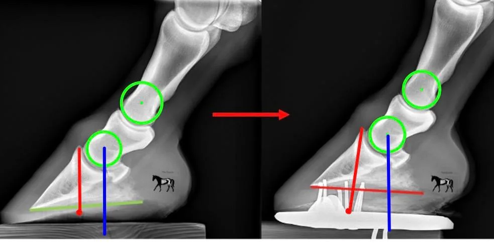
Figure X. Spatial displacement of the anatomical centre of rotation, solar reference points and the flexor moment arm following deterioration in phalangeal alignment.
These comparative lateromedial radiographs illustrate how changes in three-dimensional spatial balance alter the mechanical relationship between the distal phalanx (P3), the anatomical centres of rotation (CoR) and the structures responsible for resisting joint moments. The left image demonstrates a more favourable spatial configuration, while the right illustrates the altered relationship following deterioration in phalangeal alignment and rotation of P3.
The green circles identify the anatomical centres of rotation of the distal interphalangeal (DIPJ) and proximal interphalangeal (PIPJ) joints. The blue vertical lines represent the ground-referenced sagittal positions of the anatomical DIPJ CoR, illustrating its spatial displacement between configurations. The red lines extend from Duckett’s Dot, used here as an externally identifiable solar reference associated with the flexor apparatus, towards the palmar aspect of the digit. This provides a reproducible anatomical reference through which to visualise changes in the relationship between the distal phalanx, the flexor apparatus and the DIPJ CoR.
Two important mechanical relationships are demonstrated.
First, external solar reference points do not necessarily maintain a fixed three-dimensional relationship with the internal anatomical CoR. Although landmarks such as Duckett’s Dot can provide relatively stable references for mapping the solar surface, their spatial correspondence with internal skeletal structures depends upon phalangeal orientation. As P3 rotates and the phalangeal column becomes misaligned, the relationship between these external reference points and the anatomical joint centres changes. Consequently, apparently consistent solar mapping measurements do not necessarily imply that the underlying internal mechanical geometry has remained unchanged.
Second, rotation of P3 changes the effective moment-arm geometry around the DIPJ. In the more favourable configuration on the left, the illustrated flexor reference lies closer to the anatomical CoR in the sagittal projection. Following rotation and deterioration in phalangeal alignment, the separation between these references increases, demonstrating how changes in skeletal orientation can alter the leverage associated with the flexor apparatus. The mechanical consequences depend upon the actual tendon line of action, its perpendicular distance from the joint centre, and the external moment generated by the ground reaction force.
These changes must also be considered alongside the centre of pressure (CoP). CoP may migrate caudally relative to the solar surface while the bearing surface itself changes position relative to the skeleton. Simultaneously, rotation and displacement of the phalangeal column can alter the anatomical CoR’s position relative to the ground reaction force. Thus, compensatory CoP movement does not necessarily preserve the effective external DIPJ moment arm.
The significance for mechanical reserve is that changes in solar geometry, phalangeal alignment and CoP position are not independent loading penalties; they contribute to the resultant spatial relationships governing joint moments. Where these changes increase the effective external moment without a corresponding increase in internal leverage or other compensatory contributions, greater force is required from the structures resisting that moment.
For a flexor structure operating at a theoretical safety factor of 1.2, an additional 20% increase in tissue force would exhaust its estimated single-load reserve. This figure illustrates why relatively modest changes in spatial configuration may become important when the available mechanical reserve is already limited.
The image does not itself quantify the increase in tendon force or demonstrate a particular safety-factor threshold. Rather, it illustrates the anatomical and geometric mechanisms through which departure from ideal spatial balance can influence mechanical demand, reinforcing the need to consider external hoof proportions, phalangeal alignment and internal joint geometry together.
The central principle is that maintaining an apparently consistent solar reference does not guarantee maintenance of the same internal mechanical relationships. Spatial balance must be understood in three dimensions, with reference to both the external loading environment and the anatomical structures resisting the resulting moments.
Spatial balance and the consumption of mechanical reserve
Within the Point of Balance (PoB) framework, ideal sagittal base proportions are described as approximately 50/50 around PoB, assuming physiological three-dimensional spatial alignment.
The spatial balance concept concerns the relationship between the hoof’s bearing surface, the ground reaction force, the anatomical centres of rotation and the internal structures resisting the resulting joint moments.
In their simplest form:
Moment = Force × perpendicular moment arm
When the effective moment arm increases, the external moment increases if the applied force remains unchanged.
The opposing internal moment must increase accordingly if mechanical equilibrium is to be maintained.
For the distal interphalangeal joint (DIPJ), the deep digital flexor tendon is a principal contributor to that internal moment.
For example, if the effective external moment arm increased by 10%, with unchanged GRF and internal leverage, the required tendon force would also increase by approximately 10%.
In our theoretical Thoroughbred operating at a safety factor of 1.2, this would consume half of the original additional-load reserve.
A 20% increase would exhaust it.
This relationship is important because it provides a mechanical explanation for why spatial balance cannot be ignored or dismissed as simply a matter of external appearance or.
Changes in spatial balance alter the moment demands placed upon the distal limb, and those moments must be resisted by biological structures with finite mechanical capacity!
The precise distribution of the additional demand between tissues requires further analysis, but the underlying mechanical relationship is unavoidable.
What does the literature suggest about the sensitivity of this system?
The work of Eliashar, McGuigan and Wilson (2004) is particularly interesting in this context.
Investigating the relationship between hoof conformation and calculated force on the navicular bone in horses at the trot, the researchers demonstrated an association between distal phalanx orientation and loading of the navicular apparatus.
A figure frequently reported from this work is an approximately 4% change in peak navicular force associated with each degree of change in distal phalanx solar angle.
This deserves consideration alongside our theoretical safety-factor model.
If a comparable 4% increase per degree applied to the tensile force of a structure operating at a safety factor of 1.2, the implications would be:
Angular deterioration Hypothetical increase in tissue force Original reserve consumed
1° 4% 20%
2° 8% 40%
3° 12% 60%
4° 16% 80%
5° 20% 100%
Under those assumptions, a change of just five degrees would be sufficient to exhaust the theoretical additional-load reserve.
However, an essential distinction must be made.
Eliashar and colleagues investigated calculated navicular loading in trotting horses. The reported relationship should not be interpreted as a validated 4% increase in DDFT tensile force for every degree of DIPJ extension, nor directly extrapolated to a galloping Thoroughbred.
Navicular compressive force, DDFT tensile force, distal phalanx solar angle and combined hoof-pastern alignment are related but distinct mechanical variables.
Therefore, the table is a sensitivity illustration, not a prediction of tendon failure.
Nevertheless, the comparison is revealing.
It demonstrates how relatively small angular changes could become mechanically significant when the available tissue reserve is already narrow.
We do not need to claim that five degrees represents a universal failure threshold to recognise the importance of this possibility.
The relevant question becomes whether the changes in phalangeal alignment commonly observed alongside hoof imbalance produce sufficient additional internal demand to consume a meaningful proportion of the available reserve.
CoP compensation does not necessarily preserve the mechanical reserve.
The studies of Van Heel and colleagues add another important dimension.
During hoof growth, the centre of pressure (CoP) does not simply migrate in direct proportion to the changing external geometry of the hoof.
Compensatory changes occur.
This might initially suggest that the horse can accommodate considerable hoof growth without an equivalent increase in internal mechanical demand.
However, Moleman and colleagues demonstrated that DIPJ moments can increase during the shoeing interval despite compensatory changes in CoP position.
The explanation lies in the spatial reference used to describe movement.
CoP can migrate caudally within the solar surface while that surface itself changes position relative to the skeleton.
At the same time, alterations in phalangeal alignment may change the position of the anatomical DIPJ centre relative to the ground reaction force.
The resulting change in the external moment arm therefore depends on the net spatial relationship between the CoP and anatomical joint centre, rather than CoP movement within the hoof alone.
This gives us three important components to consider: the movement of the bearing surface relative to the skeleton, the redistribution of CoP within that surface, and the change in position of the anatomical joint centre associated with phalangeal alignment.
These are not three independent sources of additional tendon force. They are geometric contributions to the same resultant moment arm.
That distinction is essential.
Spatial displacement, angular change and CoP redistribution must be understood together because their combined effect determines the net external moment.
Where these changes increase the effective moment arm, they consume part of the same finite mechanical reserve.
This explains why two horses with apparently similar external hoof proportions may experience different internal mechanical demands.
Their pressure distribution, skeletal alignment and internal leverage may be different.
Equally, a horse may exhibit considerable external morphological deviation while compensatory mechanisms limit the increase in internal demand.
The existence of compensation does not invalidate the significance of spatial balance.
It explains why the relationship between external imbalance and internal tissue loading is not necessarily linear or universal.
Does a narrow safety factor mean a narrow tolerance for hoof imbalance?
This is the central question.
If a flexor structure has only 20% additional force capacity available under a specified loading condition, it is reasonable to suspect that its tolerance for mechanically unfavourable changes may be limited.
The Eliashar findings, together with the changes in DIPJ moments reported during hoof growth, suggest that relatively modest changes in hoof geometry and alignment can influence internal loading.
At the extreme end of equine locomotor performance, where flexor structures may already be operating with limited mechanical reserve, these changes become particularly relevant.
However, we must distinguish between two types of tolerance.
The first is the tolerance for additional tissue force.
Under our stipulated safety factor of 1.2, that reserve is mathematically defined: an additional 20% of the initial operating force.
The second is the tolerance for external spatial deviation.
That cannot be established from the safety factor alone.
For example, how far can a hoof deviate from 50/50 around PoB or how many degrees of misalignment before the relevant internal tissue demand increases by 20%?
Is the theoretical boundary reached at 55/45? At 60/40? At 5 degrees broken back? Or at a different proportion entirely?
We cannot currently assign a universal answer.
To do so, we would need to establish how changes in base proportions and phalangeal alignment influence the net CoP-to-joint-centre moment arm, GRF magnitude and direction, and internal tendon leverage.
The same external proportions may also develop through different mechanisms. Cranial hoof growth and caudal hoof collapse are not mechanically interchangeable simply because they produce similar measurements.
Consequently, the relationship between divergence from ideal spatial balance and additional tissue force may differ considerably between individuals.
A narrow mechanical reserve does not necessarily translate into a fixed or equally narrow tolerance in hoof base proportions.
But neither does the absence of a known numerical boundary mean that spatial deviation is mechanically inconsequential.
The moment demand still changes according to the resultant force geometry.
The question is how much it changes, and how much reserve the individual horse possesses to accommodate that change.
What about horses with much greater mechanical capacity?
The galloping Thoroughbred provides an interesting example because it represents a demanding locomotor environment.
But the same theoretical calculation may produce a very different result in a horse with a larger reserve relative to its operating load.
Consider two hypothetical horses exposed to the same proportional increase in tissue force.
Horse A operates at a safety factor of 1.2.
Horse B operates at a safety factor of 2.0.
If the relevant tissue force increases by 20%, Horse A’s theoretical safety factor falls to 1.0.
Horse B’s falls to approximately 1.67.
The mechanical change is identical in relative terms, but the remaining capacity is very different.
This could be relevant to horses with greater tendon cross-sectional area relative to their loading demands, different anatomical leverage, lower locomotor intensity or different tissue properties.
A larger tendon cross-sectional area may increase ultimate force capacity, assuming comparable material strength. But it does not automatically guarantee a higher safety factor, because actual operating force and tissue quality must also be considered.
Likewise, a horse travelling at a slower gait may experience a substantially different loading environment from one galloping at racing speed.
The same degree of spatial imbalance could therefore represent a relatively small proportion of the available reserve in one horse and a much greater proportion in another.
Intrinsic flexor constraint may further influence the phalangeal configuration at which equilibrium is achieved, without necessarily determining the total dynamic force experienced by the tendon.
This is particularly relevant to the proposed Low Intrinsic Flexor Constraint Phenotype (LFCP).
As discussed in the previous articles, reduced intrinsic flexor constraint may permit an alternative phalangeal equilibrium. However, it does not establish that the associated external moments are mechanically inconsequential, or that the tissues possess unlimited capacity to accommodate additional loading.
The distinction between intrinsic constraint and dynamic flexor demand remains essential.
The phenotype may influence the position at which the system achieves equilibrium, while the safety factor concerns the relationship between the resulting tissue force and the tissue’s ultimate capacity.
Individual variation may change the available tolerance, but it does not remove the mechanical consequences of altered spatial relationships.
Can we begin to calculate a theoretical range of tolerance?
I believe this is a worthwhile next step.
The starting point would be to establish the relationship between deviation from ideal spatial balance and the resulting increase in effective joint moment.
Using PoB as the spatial reference, we could quantify the departure from 50/50 base proportions and changes in phalangeal alignment and investigate the corresponding changes in CoP position, and the effective external moment arm.
We could then calculate the internal force required to resist that moment, accounting for the relevant tendon leverage and other contributions.
For a theoretical horse operating at a safety factor of 1.2, the boundary would occur when the calculated tissue force reached 120% of its initial operating value.
This would allow us to express a theoretical range of spatial balance divergence under a defined loading condition.
The process could then be repeated using different starting safety factors, anatomical dimensions, tissue capacities and locomotor demands.
Rather than producing one universal acceptable hoof proportion, such a model could reveal how working tolerance varies across different mechanical circumstances.
For example, it might demonstrate that a galloping Thoroughbred operating with very limited reserve has a relatively narrow tolerance for certain forms of spatial deviation, while a horse operating at substantially lower relative tissue demand has a wider theoretical range.
It may also reveal that some forms of imbalance consume mechanical reserve more rapidly than others, particularly where changes in external proportions are accompanied by significant phalangeal displacement or loss of caudal support.
An important qualification is that the resulting range would initially represent an ultimate-load theoretical boundary, not a clinically validated safe working tolerance.
Nevertheless, such a model would provide a starting point for quantifying the mechanical consequences of divergence from ideal spatial balance.
Why this matters beyond the Thoroughbred
The purpose of this investigation is not to suggest that every horse should be managed according to the mechanical limits of a galloping racehorse.
Quite the opposite.
The extreme example helps illustrate why we need to understand individual working tolerance.
If a horse possesses substantial mechanical reserve, it may accommodate a degree of spatial deviation without approaching its theoretical ultimate-load capacity.
Another horse, operating closer to its mechanical limits, may possess considerably less capacity to accommodate the same change.
Both remain subject to the same mechanical principles.
A change in the effective moment arm alters the moment generated by an applied force, assuming that force remains unchanged.
Changes in the required internal moment can alter tendon, ligament and other tissue loading.
Whether those changes produce meaningful increases in strain, cumulative fatigue or clinical pathology depends on the individual structures and their capacity to respond.
This distinction is important because an animal’s apparent ability to function with a particular hoof configuration does not necessarily establish that the configuration is mechanically optimal.
Nor does deviation from an ideal reference automatically establish that the horse is experiencing pathological loading.
Between those positions lies the question of tolerance.
And that tolerance cannot be understood without considering both the mechanical demands created by spatial balance and the biological capacity of the tissues resisting those demands.
Conclusion
A finite reserve, but an unknown geometric boundary
The theoretical mechanical tolerance of hoof spatial imbalance is constrained by the available safety factor of the structures resisting the resulting moments.
For a high-speed Thoroughbred operating with an assumed safety factor of 1.2, an additional 20% increase in the relevant tissue force would exhaust the estimated single-load reserve.
The angular relationships discussed by Eliashar and colleagues provide a particularly interesting illustration of how relatively small changes in spatial configuration may be associated with meaningful changes in internal loading.
Taken together, these observations raise the possibility that the theoretical tolerance for certain forms of spatial imbalance may be relatively narrow in horses operating close to their mechanical limits.
However, the magnitude of external hoof imbalance required to consume that reserve depends on the capacity of the hoof–limb system to redistribute CoP, alter phalangeal alignment and modify internal leverage.
A narrow mechanical reserve therefore does not automatically establish a fixed or equally narrow tolerance in hoof base proportions.
For the wider equine population, differences in tendon cross-sectional area, tissue properties, intrinsic flexor constraint, anatomy, posture, gait and locomotor demand may produce substantially different tolerances.
Recognising this variation does not weaken the principle of spatial balance.
It reinforces the importance of understanding the mechanical consequences of deviation from it.
The principle is not that every horse must possess identical hoof proportions, nor that every deviation from ideal balance is pathological.
The principle is that changes in the spatial relationship between the ground reaction force and the structures resisting its moments can change internal tissue demand, and those structures possess finite mechanical capacity.
Understanding how much additional demand is generated by a particular spatial deviation, and how much reserve remains available to accommodate it, may ultimately provide a more meaningful understanding of individual functional tolerance.
The question I believe we should now be investigating is:
How far can the hoof deviate from its ideal spatial reference before the associated increase in moment demand exhausts the mechanical reserve available under a specified loading condition?
And perhaps the more important question that follows:
How different is that theoretical tolerance between the galloping Thoroughbred and the wider population of horses we manage every day?
Selected references
Eliashar, E., McGuigan, M. P., & Wilson, A. M. (2004). Relationship of foot conformation and force applied to the navicular bone of sound horses at the trot. Equine Veterinary Journal, 36(5), 431–435.
Van Heel, M. C. V., et al. (2005, 2006). Investigations of hoof growth, centre-of-pressure displacement and distal limb mechanical compensation.
Moleman, M., et al. (2006). Investigation of hoof growth, pressure distribution and distal interphalangeal joint moments.
Sharp, Y. (2026). Low Flexor Constraint as a Potential Contributor to Broken-Back Hoof-Pastern Alignment in the Horse. The Equine Documentalist.
Sharp, Y. (2026). Beyond Aetiology: The Mechanical Consequences of Broken-Back Hoof–Pastern Alignment and the Limits of Functional Tolerance. The Equine Documentalist.
Scientific qualification: The 4%-per-degree relationship is discussed in secondary accounts of Eliashar et al. as a change in calculated peak navicular force associated with distal phalanx solar angle. It should be verified against the full original paper before final publication. It is not an established universal coefficient for DDFT tensile force. Similarly, the starting safety factor of 1.2 is a stipulated theoretical scenario rather than a verified universal value for the galloping Thoroughbred DDFT.

